


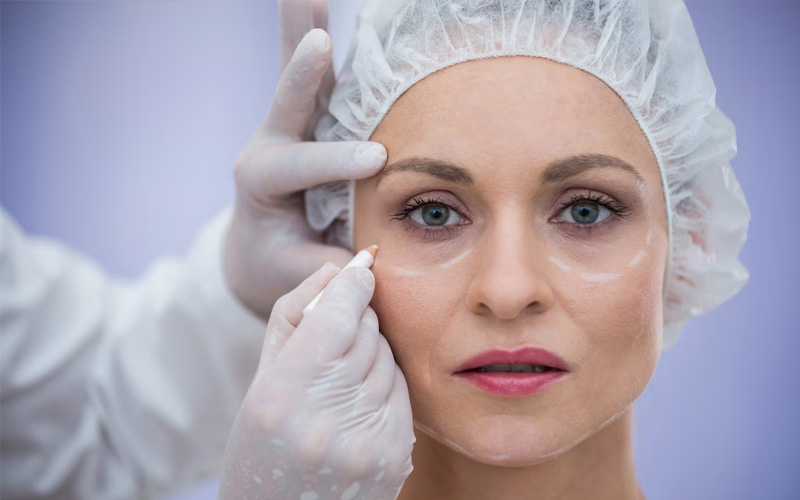


Eyelid Tumors / Eyelid Cancers
The term “tumor” refers to the presence of a lesion or mass occupying space where it shouldn’t be, anywhere in the body. These masses can be benign (non-cancerous) or malignant (cancerous). Just as in other organs, tumors can also occur in the eyes and surrounding structures, and they are relatively common. Fortunately, many of these tumors are benign growths that arise from sweat glands, sebaceous glands, eyelash follicles, and skin structures of the eyelids. However, some lesions on the eyelids can be malignant (cancers of the eye and surrounding tissues), so it is important to remain vigilant.
The most common type of malignant lesion seen in the soft tissues around the eyes, particularly on the eyelids, is basal cell carcinoma, a type of skin cancer.
Eyelid cancers are a form of epithelial tumors (skin cancers). As with all cancer types, family history and genetic predisposition play an important role in the development of cancers in the facial and periocular areas. In addition, the face is the part of our body most frequently exposed to external factors, especially UV radiation. The delicate tissues around the eyes are particularly vulnerable to these harmful external influences. Unhealthy habits like smoking, which are known to trigger carcinogenic transformations, are also significant and preventable factors. Therefore, benign and malignant tumors can frequently develop in the tissues of the face and around the eyes.
According to medical literature, papillomas are the most common benign masses found on the face, while basal cell carcinoma is the most frequently encountered malignant tumor.
Basal cells, which are found in the lower layers of the skin, are responsible for nearly 80% of all skin cancers. On the eyelids, basal cell carcinomas typically appear as raised lesions, most often on the lower eyelid. These lesions are usually poorly defined, prone to bleeding when touched, and may become ulcerated over time, resembling non-healing sores. While basal cell carcinomas on the eyelids tend to grow slowly, they can expand and deepen over time, potentially damaging the eyelids, surrounding eye structures, and even the eye itself.
Basal cell carcinomas should not be ignored, as they can progressively worsen and affect not only the aesthetic appearance but also the function of the eyes and surrounding areas. Early detection and treatment by a skilled oculoplastic surgeon are essential for achieving the best outcome.
This type or skin cancer originates from the squamous cells located on the superficial layers of the epithelium. It is rarer when compared to the basal cell carcinoma but it is much more aggressive and it can spread to surrounding tissues very fast. Mostly it starts with a precancerous form which is called “actinic keratosis”. It seen as a red nonhealing desquamated lesion on the surface.
This is the 3rd most commonly seen carcinoma of the eyelids. It is mostly seen in the middle age and older population. It is originated from the Meibomian glands which are responsible for the secretion of the outer lipid layer of the tears. It is also aggressive type of tumor which can spread quickly. Mostly seen on the upper eyelids.
Very aggressive type which spreads rapidly via blood vessels. Usually it starts as a pale brown lesion.
Lesions seen around the eye and eyelids generally require personalized treatment. Post-excision pathological examination of the lesion’s type, size, depth, and evaluation of the surgical margins will provide crucial information. Based on this, determining the best surgical approach is essential, but cosmetic and functional outcomes should not be overlooked.
The choice of surgical technique depends entirely on the lesion’s location, size, and impact on surrounding structures. For example, a lesion near the eyelashes on the upper eyelid may require minimal intervention, whereas larger lesions affecting deeper orbital tissues may necessitate a more complex approach. The primary goal is to remove the lesion completely while preserving the function of the eye and its surrounding delicate tissues. In cases where significant tissue is removed, reconstruction is performed using skin grafts or free flaps to achieve optimal results. Ensuring clear surgical margins is critical to prevent recurrence of the lesion. Grafted areas should be evaluated to ensure aesthetic acceptability, allowing patients to return to daily life with minimal cosmetic compromise. For residual lesions, treatments like cryotherapy or radiation therapy may also be considered.
One of the most important considerations in these cases is meticulous planning and care. If excision is incomplete or if critical structures such as tear ducts, eyelids, or surrounding tissues are damaged, it can lead to complications that could affect function and appearance. Early and appropriate intervention is key to achieving successful outcomes and a smooth recovery for the patient.